
KRF Clinical Practice Guidelines in Keloid Disorder (KRF Guidelines®) Intra-Lesional Chemotherapy Version 1.2018
Michael H. Tirgan, MD
BACKGROUND
Intra-lesional chemotherapy (ILC) using 5-fluorouracil (5-FU) for treatment of keloid lesions was first introduced by Fitzpatrick in 1989 [1]. In a survey conducted by the author [2], only 4.23% of keloid patients reported having received ILC injections for treatment of their keloids. In the United States, and perhaps globally, the most commonly used chemotherapy drug is 5-FU.
In preparing 5-FU for ILC, Fitzpatrick (1999) reported using 50 milligrams (mg) per milliliter (ml) concentration of 5-FU [1], which is the usual concentration of the undiluted drug in its vial [3]. Although Fitzpatrick capped the total administered dose of 5-FU at 100 mg per treatment session, he did not provide a rationale for choosing this dose or for the concentration of 5-FU used in his experiment. However, this original work might have been the reason behind the common practice of using the undiluted solution of 5-FU, i.e. the highest possible concentration of the drug for intralesional injections. The author has personally interviewed several practitioners who use 5-FU at the highest concentration of 50 mg/ml – straight out of the vial – a practice that is in line with using Kenalog-40 and injecting undiluted triamcinolone into the keloid tissue [4,5].
The author is unaware of any studies to determine the proper dosage of 5-FU for intra-lesional treatment of keloids. Other chemotherapy drugs, such as bleomycin and mitomycin, have also been used in treating keloids with variable results [6].
Indications
Readers shall consult with KRF Guidelines on Treatment Strategy as well as site specific Guidelines for the proper and up-to-date course of treatment for their patients.
ILC is most appropriate for treatment of papular, linear, or flat keloids and should be used only as a second or laterline of treatment after documenting the failure of previous treatment with intra-lesional triamcinolone (ILT). ILC should not be used as a first line treatment in any setting. In choosing ILC, the treating physician must obtain proper treatment history from the patient and affirmatively establish that ILT has already been used, at least twice, and that the keloid has either failed to respond or has progressed with ILT.
Syringe / Needle Gauge
The author recommends a Gauge 30 needle for injection of ILC. A larger gauge needle will introduce a new injury to the keloid, which may result in reactivation and worsening of the keloid lesion [5]. Figure 1 depicts the type of syringe and the recommended needle.
The 1 ml syringe, like the one depicted in Figure 1, is graduated to 1/10 and 1/50 of ml. When the syringe is filled to its last line marked as 100, it will contain 1 ml of fluid. Each graduated line on the syringe equates to 2/100 of a ml.

Figure 1. A 1 ml syringe with gauge 30 needle incorporated in the body of the syringe. This is the proper type of syringe for ILC injections. Each
graduated line equates to a volume of 2/100 of ml.
Injection Pain
ILC injections can be somewhat painful, especially when given to numerous keloids in one session. Therefore, injection pain must be addressed with all prospective patients. Informing patients about injection pain reduces the anxiety associated with first-time injections.
Slow and gentle injections as opposed to rushed and quick injections can also help to reduce discomfort. For patients who have had previous ILC injections, discussion of the pain from past injections will guide the treating physician in using methods that will reduce the pain and anxiety of subsequent injections.
Technique and Volume of Injection
The keloid lesion must be constantly and carefully inspected during the ILC injection. The goal of the injection is to deliver a minimal yet sufficient amount of the prepared solution of chemotherapy into the core of the target lesion.
Using the syringe shown in Figure 1, the needle is gently inserted into the body of the keloid. The content of the syringe is then gently injected into the keloid to the point of inducing a minimal swelling and blanching of the lesion. At this point, the needle is withdrawn. The volume of each injection, and the number of injections into a given lesion, depends on the size and thickness of the lesion.
The total dose of ILC needed for lesions that measure up to one centimeter in diameter can usually be delivered in one injection. When the lesions are linear and long, or large and flat, several injections are needed to deliver the ILC to whole or most of the lesion. A bandage shall be placed over the injected keloid for approximately one hour.
Assessment of the Efficac y of ILC
All keloid lesions must be photographed prior to the initiation of any form of treatment, including treatment with ILC, and also at every follow up visit. A pre-treatment photograph will serve as a reference point to determine the treatment’s efficacy.
Upon initiation of ILC, the injected lesion(s) must be inspected within three to four weeks, at which point the patient and the provider will together determine whether ILC was effective.
If ILC is determined to have been effective, it can be repeated during the follow-up session. Injections should be repeated every three to four weeks until the maximum response is achieved, at which point the treatment shall be halted and the lesions shall be photographed and observed.
In contrast to ILT, ILC can be a curative treatment. New and small keloid lesions may totally disappear after one or two ILC injections. Lesions that respond to ILC should be monitored for potential recurrence.
When used in patients with numerous lesions, some lesions will respond better than others. When a lesion achieves a good response, it should no longer be treated. Patients should be advised to monitor their lesions and to return in the event of a recurrence.
If ILC is thought to be ineffective after the first injection, it is reasonable to repeat the injection, with the same drug and the same concentration of chemotherapy. If after two consecutive injections the patient and the physician determine that the particular drug used in ILC was ineffective, the treatment with that drug must be abandoned and another chemotherapy drug should be utilized. There is no justification to either use higher doses of ILC or to continue with a treatment that does not show efficacy after one or two injections. This practice will only delay the treatment course.
h3>Frequency of Injections
When effective, ILC injections often need to be repeated until maximum response is achieved. Most patients who observe improvement in their keloid lesions often report that the ILC’s efficacy is long-lasting.
In patients who do respond to treatment and need repeated injections to maintain their benefit from ILC, a determination must be made as to the longevity of the efficacy, which varies from patient to patient. Future injections must be given at intervals that match the longevity of the benefit from ILC. The author has encountered several patients who only need maintenance ILC injections once every few months.
RECOMMENDED DRUGS
In choosing chemotherapy drugs for treatment of a benign disease, only drugs that are non-carcinogenic, nonmutagenic, and with a reasonable short- and long-term safety profile should be used. Among the numerous, available chemotherapeutic drugs, the author recommends the following three drugs for intralesional treatment of keloids:
- 5-FU
- Vincristine
- Docetaxel
These drugs must be used in sequence, i.e. 5-FU should be used first, and if the keloid lesions show resistance to this drug, then vincristine shall be used. Docetaxel shall only be used in lesions that have failed to respond to both 5-FU and vincristine. This sequence is derived from safety, side effect profile, and the complexities involved in proper administration of each one of the three drugs.
General Precaution – Embryo-Fetal Risks
Although very low doses of chemotherapy drugs listed above will not pose any risks to adult patients, pharmaceuticals that are genotoxic and target rapidly dividing cells are all presumed to be teratogenic and/or lethal to an embryo/fetus.
In female patients, the growth and maturation phase of folliculogenesis (four to six months) is most susceptible to persisting deoxyribonucleic acid (DNA) damage and may potentially result in embryo-fetal malformations.
In male patients, the chemotherapy drugs may cause DNA damage in sperm, potentially resulting in adverse effects in the conceptus with a female sexual partner.
To minimize the risk of adverse embryo-fetal effects, all patients – male or female – shall be warned about the potential embryo-fetal risks of ILC and advised to use contraception
for a period of six months after cessation of therapy.
5-Fluorouracil
When considering ILC to treat keloid lesions, 5-FU shall be the first drug used. In the author’s experience, approximately 30% of keloid lesions respond to this treatment.
Recommended Dosage
When used in treating cancers, 5-FU is administered intravenously, often as a continuous infusion at a dose of 1,000 mg per metered squared (m2) body surface area per day for four to five days. But, 5-FU has a very short halflife in the blood. The effective cytotoxic concentration of the drug that is achieved inside tumor tissue is measured in micrograms (mcg) per gram of tumor mass [7]. To the author’s best knowledge, the cytotoxic concentration of 5-FU needed to induce a response in keloid lesions has never been studied.
As a matter of principle, it is most logical to always use the lowest effective dose of any medication for treatment of any disease. ILC and keloid disorder are no exception to this principle. Therefore, when injecting keloids with triamcinolone or chemotherapy drugs, the lowest effective dose of these drugs should be used.
Fitzpatrick (1999) proposed a dose of up to100 mg of 5-FU per treatment session [1]. Compared with standard oncology doses, this dose may appear to be low and safe. However, some oncology patients exhibit severe systemic side effects to 5-FU. Since the late 1980s, there has been an increasing number of case reports describing severe toxicity (including death) from treatment with 5-FU among oncology patients. This toxicity results from deficiency of dihydropyrimidine dehydrogenase (DPD), which is a key enzyme in the metabolism of 5-FU [8].
The disposition and pharmacokinetics of 5-FU were evaluated in one of the first reported DPD-deficient patients [8]. A test dose of 5-FU at 25 mg/m2 was administered to this patient as an intravenous bolus, after which plasma, urine, and cerebrospinal fluid were sampled at specified times. The 5-FU was detected in the plasma at unusually high levels for ≥ 8 hours, demonstrating a markedly altered pharmacokinetic pattern for this patient and a biologic halflife of 159 minutes, in contrast to a group of 10 normal control patients who were given a typical dose of 5-FU (450 mg/m2) in which the half-life of the drug was 13 minutes ± 7. The test dose used in this experiment was lower than the dose schedule proposed by Fitzpatrick [1].
The emergence of more detailed understanding of various mutations in the genes that encode the production of DPD has resulted in better understanding of the potentially severe toxicity associated with 5-FU [9].
Therefore, in the absence of proper dose response studies, and to prevent potential 5-FU toxicity in DPD deficient subjects, it is of the utmost importance to use a very low dose of 5-FU in treating keloid lesions. In preparing 5-FU for intralesional injections (IL-5FU), the author recommends a maximum dose of 1 mg per treatment session, which will easily achieve a cytotoxic concentration of 5-FU [7] in the keloid tissue. This dose shall be added to the standard preparation of triamcinolone for ILT injection (see KRF Guideline – ILT).
Preparation of the 5-FU Solution for Injection
5-FU is commercially available in 10 ml or larger size vials at a standard concentration of 50 mg/ml. The author uses the following steps in preparing IL-5FU:
- 5-FU is first drawn into a 1 ml syringe (Figure 1) to the first graduated line. Each graduated line represents a volume of 2/100 ml and will contain 1 mg of 5-FU.
- Triamcinolone is then added to 5-FU in the same syringe by drawing 2/10 ml of Kenalog-10, which contains 2 mg of triamcinolone (see KRF Guideline – ILT).
- Normal saline is then drawn into the same syringe to a total volume of 1.0 ml.
This mixture will contain 1 mg of 5-FU and 2 mg of triamcinolone. Each graduated line of the mixture will contain 20 mcg of 5-FU and is ready for injection into the target keloid lesions.
Side effects
When injected properly, IL-5-FU is well tolerated and does not cause any systemic or local side effects at the injection site. Repeated ILC injections using 5-FU, vincristine, or docetaxel, can all result in hyperpigmentation at the injection sites (Figure 2) in patients with dark skin color, Fitzpatrick skin type IV-VI.
Vincristine
Vincristine is the second drug of choice for the intra-lesional treatment of keloid lesions. It should only be considered, however, for treatment of lesions that have failed to respond to IL-5FU. In the author’s experience, approximately 60% of keloid lesions respond to this treatment.
The standard method for the administration of vincristine in oncology is intravenously; however, intra-lesional vincristine (IL-VCR) has been used in the treatment of Kaposi’s sarcoma lesions [10].
Extravasation of vincristine is known to cause severe tissue necrosis [11]. IL-VCR harnesses this exact property of vincristine for treatment of keloids with the aim of inducing a controlled tissue necrosis within the keloid lesion. Figures 3-5 depict the powerful cytotoxic activity of IL-VCR and its ability to induce tissue necrosis in a tumoral keloid (Figure 3). The lesion was injected with 10 mcg of vincristine. Clinically, noticeable tissue necrosis was seen in three weeks (Figure 4). Further tumor shrinkage was achieved with repeated IL-VCR injections (Figure 5). This case provides proof of concept that significant tumor kill can be achieved in a keloid tumor with microgram doses of vincristine. In the absence of a properly performed dose response study, the evidence provided in this one case can be used as guidance to estimate the amount of vincristine needed for much smaller keloids.
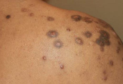
Figure 2. Typical ILC-induced hyperpigmentation.

Figure 3. Tumoral keloid of right shoulder area in a 27-year-old African American male.
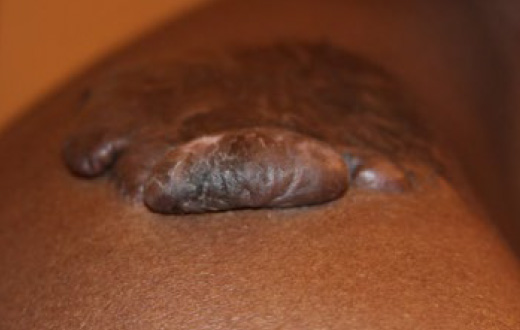
Figure 4. The same keloid, 21 days after a single injection of 10 mcg of IL-VCR. The keloid displays signs of tissue necrosis and volume loss. A second IL-VCR, 10 mcg, was injected during this visit.

Figure 5. The same keloid, 42 days after the first injection, showing significant necrosis and volume loss from the two previous IL-VCR injections.
Recommended Dosage
In preparing vincristine for intra-lesional injections, the author recommends a very low dose of this drug, no more than 20 mcg of vincristine, to be added to a standard preparation of triamcinolone for ILT injection, with final volume of 0.4 ml of the solution that is to be used for injection.
Extreme care must be given to choose and prepare the correct dosage of vincristine in the smallest possible volume in order to minimize the chance of causing damage from vincristine to the normal surrounding tissues. Vincristine is a very potent vesicant and if injected incorrectly will result in serious tissue necrosis.
Recommended Volume of Injection
Vincristine is commercially available in 1-2 ml vials at a concentration of 1 mg/ml. This concentration is equivalent to 1,000 micrograms of the drug per ml. Extreme care must be exercised to extract the correct amount of the drug into the syringe that is to be used for IL-VCR injection. The dose and volume of vincristine to be injected into the keloid lesion depends on the size of the lesion. Table 1 provides a guidance for choosing proper dose and volume of vincristine.
Table 1. Recommended dose and volume of vincristine for intralesional injection of keloid lesions.

Abbreviations: mm=millimeter, ml=milliliter
Preparation of the Solution for Injection
The author uses the following steps in preparing IL-VCR:
- Vincristine is first drawn into a 1 ml syringe (Figure 1) to the first graduated line. Each graduated line represents a volume of 2/100 ml and will contain 20 mcg of vincristine. This is the maximum amount of vincristine that should be used to inject keloids in a single treatment session.
- Triamcinolone is then added to the same syringe by drawing 1/10 ml of Kenalog-10, which contains 1 mg of triamcinolone (see KRF Guideline – ILT).
- Where available, liposomal bupivacaine [12] or normal saline is then drawn into the same syringe to increase the total volume to 0.4 ml.
This mixture will contain 20 mcg of vincristine and 1 mg of triamcinolone. Each graduated line of the mixture will contain 1 mcg of vincristine and is ready for injection into the target keloid lesions.
Side effects
IL-VCR causes two distinct side effects:
Hyperpigmentation is a common side effect of all drugs used for ILC / IL-VCR, especially when used repeatedly in individuals with dark skin color and African Americans (Figure 2).
Post injection neuropathic pain at the injection site is the most important and unique side effect of IL-VCR. This pain usually starts 24-28 hours after the injection and will last four to seven days. The intensity and the duration of the pain is directly related to the injected dose of vincristine and to some extent to the location of the treated keloid. The same dose of IL-VCR will cause a much more severe pain in a centrally located anterior chest keloid when compared with a shoulder keloid.
Proper pain management is the key factor in gaining patient compliance to IL-VCR. All patients must be educated about the expected pain and prepared to properly manage this pain. Where available, liposomal bupivacaine should be added to the mixture of the IL-VCR solution. This drug can provide up to 72 hours of local anesthesia. Once the anesthetic effect wears off, the patient can either take oral non-steroidal antiinflammatory drugs (NSAIDs) or return to the clinic for a second injection of liposomal bupivacaine. When this drug is unavailable, NSAIDs should be used for management of mild to moderate pain and when needed, oral opioids should be used for more severe pain.
Because IL-VCR at doses of over 5 mcg can cause severe pain, the physician must maintain an open line of communication with all patients so that the pain can be properly managed.
Pain associated with doses of 1-2 mcg is usually minimal and may be perceived as a mild burning sensation or the feeling of running electric current at the injection site. At doses above 5 mcg, pain can become quite bothersome, especially at night.
It is complex dosing and the treatment induced pain that ranks IL-VCR second after IL-5FU. Table 2 provides a summary for the level of pain associated with different dose levels of vincristine along with the recommended interventions for pain control.
Table 2. Pain associated with IL-VCR and the recommended pain management

Abbreviations: NSAID=non-steroidal anti-inflammatory drugs.
Treatment of Large Keloids
When treating a very large keloid with IL-VCR, especially one centrally located in the anterior chest, the treating physician must first test the efficacy of the treatment by injecting a small portion of the keloid that is no wider than 20 mm with a test dose of no more than 5 mcg.
The efficacy of this treatment shall then be assessed in two to three weeks. Once proven effective, IL-VCR can then be injected into the rest of the keloid, but in a segment by segment manner. Treatment of an anterior chest keloid that measures 10 cm in diameter should be divided in at least three sessions, with one-third of the lesion to be injected in each session.
Docetaxel
Docetaxel is the author’s third choice of chemotherapy drugs for treatment of keloid lesions. Intra-lesional docetaxel (ILTAX) is used only in patients who have failed to respond to both IL-5FU and IL-VCR. In the author’s experience, approximately 70% of lesions respond to this treatment. New formed keloids respond very well to this treatment.
Recommended Dosage
Docetaxel is available in 1 ml or 4 ml vials at a concentration of 20 mg/ml. In preparing docetaxel for ILC injections, the author recommends a very low dose of this drug to be added to the standard preparation of triamcinolone for ILT injection, with no more than 400 micrograms of docetaxel per 1.0 ml of the final preparation that is to be injected.
Preparation of the Solution for Injection
The author uses the following steps in preparing IL-TAX:
- Docetaxel is first drawn into a 1 ml syringe (Figure 1) to the level of the first graduated line on the syringe. Each graduated line represents a volume of 2/100 ml and will contain 400 mcg of docetaxel.
- Triamcinolone is then added in the same syringe by drawing 2/10 ml of Kenalog-10, which contains 2 mg of triamcinolone (see KRF Guideline – ILT).
- Normal saline is then drawn into the same syringe to a total volume of 1.0 ml.
This mixture will contain 400 mcg of docetaxel and 2 mg of triamcinolone. Each graduated line of the mixture will contain 8 mcg of docetaxel and is ready for injection into the target keloid lesions. The amount of docetaxel that is to be injected into the keloid lesion depends on the size of the lesion.
Side effects
IL-TAX causes two distinct side effects:
Hyperpigmentation is a common side effect of all drugs used for ILC / IL-TAX, especially when used repeatedly in individuals with dark skin color and African Americans (Figure 2).
Mild to moderate post injection pain at the injection site may be experienced when a large lesion is injected with doses of over 200 mcg. NSAIDs are usually sufficient for pain control from IL-TAX. The pain from IL-TAX is significantly less than the neuropathic pain of IL-VCR.
Acute Hypersensitivity Reaction is the most important side effect of docetaxel. Both the docetaxel and the solvent in which it is dissolved (polysorbate 80) can contribute to acute hypersensitivity reactions [13]. When used in treating cancer patients, despite proper pre-medications, this hypersensitivity reaction can manifest itself within minutes of intravenous infusion of the drug.
In the author’s experience with off-label use of IL-TAX in treating keloids, he noticed a mild reaction of possible hypersensitivity in three of his first 15 patients. One patient experienced flushing and redness on the face, a second patient felt slightly lightheaded, and the third patient experienced a metallic taste in his mouth.
The reactions in all three cases was immediate and occurred while the drug was still being injected into the keloids. The procedure was immediately abandoned and all three patients recovered fully in matter of few minutes.
These events prompted the author to empirically premedicate all subsequent patients. After experience with more than 50 premedicated patients, the author has not witnessed a case of hypersensitivity reaction.
Mandatory Premedication Regimen
Oral dexamethasone at the dose of 8 mg twice per day should be started on the day before the planned injection of IL-TAX and continued on the day of injection.
Test Dose Injection
A test dose must first be injected before injecting the whole planned dose of IL-TAX. A minute amount of prepared solution, a volume of less than 1/100 ml, shall be injected inside the target lesion. The patient shall then be observed for approximately five minutes. Absent any hypersensitivity reaction, the injection can be resumed and the rest of the planned dose gradually injected into the keloid. The patient shall be monitored for signs of hypersensitivity while the drug is being injected.
Delayed Hypersensitivity Reaction to docetaxel is another known side effect of docetaxel in oncology patients. The author has seen two cases of delayed cutaneous hypersensitivity reaction to IL-TAX. One patient, despite the proper use of premedication, noticed mild itching at the drug’s injection site. The itching started a day after the injection and lasted for a few days. When re-treated with docetaxel, the patient experienced similar symptoms, but this time the itching occurred considerably sooner. Another patient developed a localize rash at the sites of injection within days after the injection. The same rash occurred within 24 hours after the next IL-TAX injection. Treatment with docetaxel was abandoned in both patients.
The occurrence of delayed hypersensitivity reaction should prompt termination of IL-TAX treatment in all such patients.
Case Study 1
A 29-year-old male presented with a tumoral anterior chest wall keloid (Figure 6). This keloid had been present for approximately 13 years. The patient reported no previous treatments.
The keloid was first treated with ILT, with minimal response after three injections. The keloid was then treated with a combination of ILT and cryotherapy for which a partial response was achieved (Figure 7). At this point, decision was made to switch to ILC. Near complete remission was achieved and maintained using intermittent doses of ILVCR at 2 mcg every few months.

Figure 6. Central chest keloid in a 29-year-old male (April 2014).
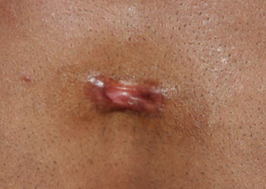
Figure 7. The same patient prior to initiation of ILC. Notice the reduction in the mass of keloid from prior ILT and cryotherapy (September 2014).

Figure 8. The same patient after six doses of ILC. Note the significant reduction in the mass (September 2015).

Figure 9. The same patient after several ILC injections (July 2018).
References
- Fitzpatrick RE. Treatment of inflamed hypertrophic scars using intralesional 5-FU. Dermatol Surg. 1999 Mar;25(3):224-32.
- Tirgan, MH. Web Based Investigation of Natural History of Keloid Disorder, an Online Survey.
- 5-Fluorouracil, package Insert.
- Tirgan, MH. Clinical Practice Guidelines in Keloid Disorder – Intralesional Triamcinolone.
- Tirgan, MH. Intra-lesional Steroid Injections: Can they be harmful to some patients? Results of an online survey. International Journal of Keloid Research. 2017 vol. 1 no. 1, July 1, 21-28.
- Jones, CD et al. The Use of Chemotherapeutics for the Treatment of Keloid Scars Dermatol Reports. 2015 May 21; 7(2): 5880.
- Zheng JF, Wang HD. 5-Fluorouracil concentration in blood, liver and tumor tissues and apoptosis of tumor cells after preoperative oral 5’-deoxy-5-fluorouridine in patients with hepatocellular carcinoma World J Gastroenterol. 2005 Jul 7; 11(25): 3944–3947.
- Ezzeldin H, Diasio R. Dihydropyrimidine dehydrogenase deficiency, a pharmacogenetic syndrome associated with potentially lifethreatening toxicity following 5-fluorouracil administration. Clinical colorectal cancer. 2004;4(3):181-189.
- Diasio RB, Beavers TL, Carpenter JT. Familial deficiency of dihydropyrimidine dehydrogenase. Biochemical basis for familial pyrimidinemia and severe 5-fluorouracil-induced toxicity. J Clin Invest 1988; 81:47-51.
- L. Brambilla, et al. Intralesional vincristine as first-line therapy for nodular lesions in classic Kaposi sarcoma: a prospective study in 151 patients. British Journal of Dermatology v162 n4 (April 2010): 854-859.
- R. A. Ener, S. B. Meglathery, M. Styler. Extravasation of systemic hemato-oncological therapies Annals of Oncology, Volume 15, Issue 6, 1 June 2004, Pages 858–862.
- Liposomal bupivacaine, package insert.
- Docetaxel, package insert.


