Worsening of Keloids During Pregnancy:
Results of an online survey
Michael H. Tirgan, MD
ABSTRACT
Background
Physiological changes during pregnancy have a significant impact on many organ systems including the skin. Many skin conditions, including keloids, are known to worsen during pregnancy. Although worsening of keloids during pregnancy has been reported in the literature, the actual rate and incidence of this phenomenon have not been previously reported.
Objective: To evaluate the relationship between pregnancy and change in the rate of growth or symptomatology of keloids among women who had at least one keloid lesion prior to becoming pregnant.
Material and Methods: An online survey was launched in November 2011 asking participants to provide answers to questions about their keloids. Female patients were asked about the potential impact of pregnancy on their keloids. Descriptive statistics are provided.
Results: As of February 13, 2022, 1886 individuals had participated in this survey, and 1715 provided information about their gender (male, 561 [32.71%]; female, 1154 [67.29%]). Among women, 297 reported that they had developed their first keloid prior to becoming pregnant and only 19% of them reported significant worsening of their keloids during pregnancy.
Conclusions: To the author’s knowledge, this is the first systematic review and report of the incidence of worsening of keloids during pregnancy. The cause of the worsening of keloids during pregnancy is directly related to the factors that are inherent to the keloid tissue and not the pregnancy itself.
INTRODUCTION
The physiological effects of pregnancy can affect common skin conditions [1]. However, the effect of pregnancy on keloid disorder (KD) has not been systematically studied. The first published report of pregnancy impacting the natural history and biology of KD appears to be by Jacobsson (1947) who described the development of a keloid during pregnancy in a facial scar that had been dormant for 4 years [2].
Mustafa et al. (1975) reported a case of a young female patient who had been struggling with bilateral ear keloids since she was 12 years old [3]. The patient had undergone multiple surgeries, each time requiring a wider excision. At age of 22, during the fourth month of her first pregnancy, her right ear keloid started to rapidly grow. By the seventh month into her pregnancy, she developed a massive keloid on her right ear. More recently, Ibrahim et. al. (2020) reported two cases of extreme worsening of keloids during pregnancy [4].
In the present study, the rate and causality of the worsening of KD during pregnancy were evaluated by an online survey about different aspects of KD, including the impact of pregnancy on the natural history of KD. The present study reports on the self-assessments of patients who completed the survey.
MATERIALS AND METHODS
A comprehensive questionnaire was developed to survey a large cohort of consecutive unselected patients with KD. The study was initially approved in November 2011 by the IRB of St. Luke’s Roosevelt Hospital in New York. The study was subsequently transferred to Western IRB.
Patients with KD who were interested in participating in the study were provided with access to the study questionnaire at www.KeloidSurvey.com. Adult participants provided their informed consent electronically.
The survey included age, ethnic background, family history, extent and distribution pattern of the keloid lesions, history of pregnancy in females, and how pregnancy might have impacted the behavior of KD.
Participants’ access to the survey tool was limited to one access per computer IP address. They could skip a question if it did not apply to them, or if they did not wish to answer. The study dataset was accessed on February 13, 2022. Descriptive statistics and the rates of patient reported events are presented. The current study is based on the analysis of data provided by this cohort of women (n = 297) who already had a keloid prior to becoming pregnant.
RESULTS
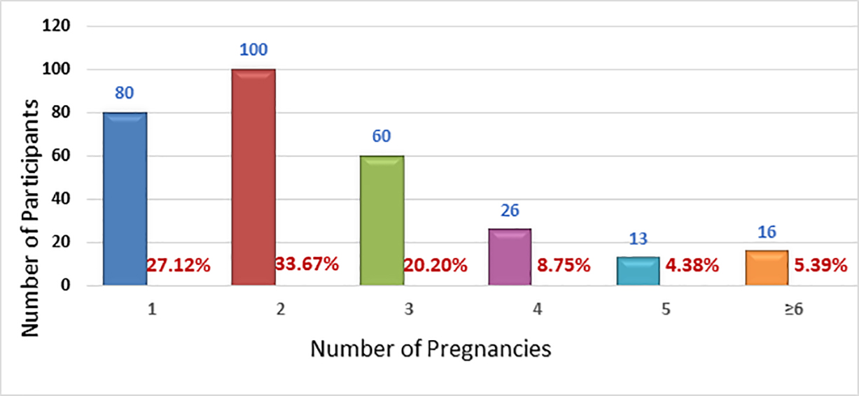
The study was opened for accrual on November 14, 2011. As of February 13, 2022, 1886 individuals had participated in this survey, 1715 individuals provided information about their gender (male, n = 561 [32.71%]; female, n = 1154 [67.29%]). Among the female participants, 916 provided an answer as to whether they have ever been pregnant. Three hundred ninety-six female participants (396) indicated that they had become pregnant at some point prior to taking the survey and provided answers to the pregnancy-related questions. Participants were asked whether they had developed their first keloid before, during, or after they became pregnant. Nineteen participants reported that their first keloid developed during their pregnancy and 297 reported that they had developed their first keloid prior to becoming pregnant. The current study is based on the analysis of data provided by this cohort of women (n = 297) who already had a keloid prior to becoming pregnant. Figures 1 and 2 depict the number of pregnancies, and deliveries reported by this cohort of women who reported having developed their first keloid prior to becoming pregnant. Approximately 33.67% of participants reported two prior pregnancies, 27.12% only one prior pregnancy, and 20.20% reported three pregnancies prior to taking the survey.

FIGURE 1: Number of pregnancies reported by participants (n = 295).

FIGURE 2: Number of deliveries reported by participants prior to taking the survey. Most participants had up to three term pregnancies and delivery. It is natural that some women might have become pregnant and had a miscarriage (n = 275).
Age
Participants were also asked to provide their age at the time they took the survey; 295 participants provided an answer. Figure 3 depicts participants’ ages at the time they took the survey.

FIGURE 3: Participants’ age at the time they took the survey (n = 295).
Age of onset/clinical presentation of keloid disorder
Participants were also asked to provide their age at the time they developed their first keloid lesion. A total of 295 participants indicated their age at the time they developed their first keloid lesion. The peak age of onset was 12 years; 258 participants (87.46%) had developed their first keloid between the ages of 5 and 25 years.
A total of 215 participants (72.88%) were between the ages of 1 and 17 when they developed their first keloid, making KD a predominantly pediatric illness. Figure 4 depicts participants’ reported age at the time they developed their first keloid lesion.

FIGURE 4: Age of onset/clinical presentation of keloid disorder among the study population (n = 295).
Country of Birth
A total of 295 participants provided information about their country of birth. The majority of participants (66.78%) were born in the United States, 4.41% were born in India, 4.07% in the Philippines, and 2.71% in the United Kingdom. Figure 5 depicts the country of birth of the study participants.
Although this distribution pattern correctly represents the country of birth of those who participated in this study, it is by no means a true reflection of the epidemiology of KD. Lack of access to the internet in many regions worldwide, as well as the fact that the survey has been available only in English, limits the availability of the survey to many keloid patients.

FIGURE 5: Country of birth of the study participants (n = 295).
Ethnicity
Participants were asked to provide their ethnic background. A total of 296 participants indicated their ethnicity: 42.23% identified as African American, 16.89 as White/North American, 7.09% as Hispanic, and 5.07% as far East Asian.
Pattern of Distribution of Keloid Lesions
Participants were asked to provide detailed information about the distribution of the keloid lesions throughout their skin. A total of 291 participants provided this information. Participants who had multiple keloids in different parts of their skin were asked to indicate all sites of disease involvement. The shoulder, upper chest, upper arm, pelvis, earlobe, lower chest, and neck were the most frequently involved area of the skin among the study participants. Figure 6 depicts the distribution patterns of keloid lesions among the study participants.

FIGURE 6: Pattern of distribution keloid lesions among study participants (n = 291).
Morphology of Keloid Lesions
Participants were asked to describe the shape and appearance of their keloid lesion(s). To facilitate this, a reference image guide was provided online. A total of 297 participants provided this information. Participants who had multiple keloids with different morphologies were asked to describe each type of keloid.
Nodular morphology was reported in 63% of participants followed by linear keloids in 57% and flat keloids in 43% of the participants. Approximately 34% of patients considered their keloids to be massive, with keloid lesions occupying large areas of their skin. Figure 7 depicts the distribution patterns of the shape of the keloid lesions among the study participants.

FIGURE 7: Morphology of keloid lesions among study participants (n = 297).
Triggering factors
Participants were asked to provide information about the factors that triggered the formation of their keloids; 283 participants answered this question. Figure 8 shows the frequency of various triggering factors within this population. Many patients reported more than one triggering factor.

FIGURE 8: Triggering factors reported by participants (n = 283).
Number of Keloids
Participants were asked to provide information about the number of keloid lesions that they had at the time they took the survey. A total of 292 participants provided this information. Figure 9 depicts the distribution pattern of the number of keloid lesions that each participant reported within this population. Approximately 25% of the participants reported having 10 or more keloid lesions.

FIGURE 9: Distribution pattern, number of keloid lesions in each participant (n = 292).
Impact of pregnancy on rate of growth of keloid lesions
The impact of pregnancy on the rate of growth of keloid lesions was assessed by asking participants to provide only one answer to one set of multiple-choice questions that would explore observed impact of the pregnancy on the behavior of the keloid lesions. 273 participants answered the questions about impact of their pregnancy on behavior of their keloids. Approximately 57% of participants in this study reported that pregnancy has no impact on the rate of growth of their keloids, 23% reported minor impact on the rate of growth of their keloid during pregnancy, 11% reported moderate worsening and only 8% reported significant worsening of their keloids during pregnancy.

FIGURE 10: Impact of pregnancy on the rate of growth of keloid lesions (n = 273).
Impact of pregnancy on keloid-related symptoms
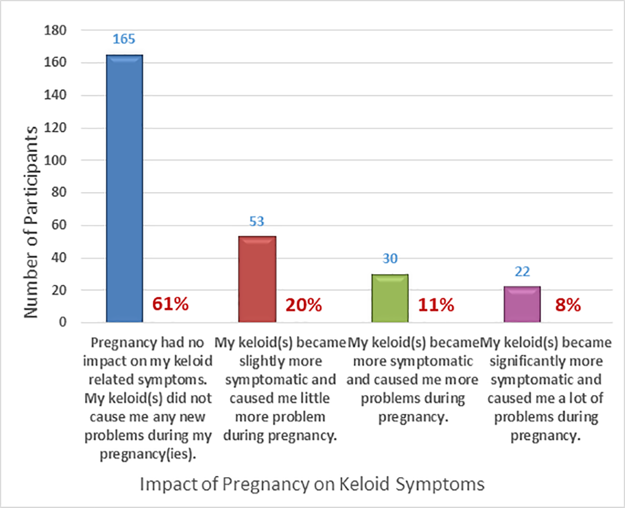
The impact of pregnancy on the symptoms associated with keloid lesions was assessed by asking participants to provide only one answer to one set of multiple-choice questions that would explore participants perception of the impact of the pregnancy on the symptoms of their keloids. Figures 11 depict the responses by the 270 participants who answered the questions about impact of their pregnancy on behavior of their keloids during pregnancy. Sixty one percent of women reported that their pregnancy had no impact on their keloid symptoms. Twenty percent of participants reported slightly more symptoms, 11% reported moderate increase in their symptoms, with only 8% reporting significant worsening of keloid related symptoms during pregnancy.

FIGURE 11: Impact of pregnancy on keloid-related symptoms (n = 270).
Timing of worsening of keloids during pregnancy
Figure 12 depicts the responses by the 141 participants who answered the questions about the time when they noticed the most changes of their keloid during their pregnancy: 15.60% reported that they noticed the most impact during the first trimester of their pregnancy. 34.04% during the second trimester, and 50.35 % during the third trimester of pregnancy.

FIGURE 12: Timing of most impact of pregnancy on keloids (n = 142).
DISCUSSION
Pregnancy can affect the natural history of several illnesses, but its true impact on the behavior and biology of KD has not been studied. To the author’s knowledge, this is the first systematic study of the impact of pregnancy on biology and clinical behavior of KD.
Subset analysis was performed to identify the factors that might have been associated with more severe impact of pregnancy. However, the sample size in the two cohorts who reported moderate to severe worsening of the keloids was not sufficient to draw meaningful conclusions.
Although the correlation between worsening of keloids and pregnancy has been reported as case reports since 1947, this causal association has never been formally studied. In their recent case reports, Ibrahim et al. contributed this effect of pregnancy to the hormonal effects of pregnancy on the keloid tissue.
In the present study, it was hypothesized that the association between pregnancy and worsening of keloids is significantly more complex, in that the hormonal changes during pregnancy are generally the same among all women who become pregnant and carry their pregnancy to term. However, the results of the present study suggests that the impact of pregnancy on keloids is not the same among all pregnant women. In particular, 57% of participants in this study reported that pregnancy did not have any impact on the rate of growth of their keloids and 33% reported minor impact on the behavior of their keloids.
Thus, all women in various cohorts of this study may have similar pregnancy-related hormonal changes, indicating that the variable among these women resides in the keloid tissue and factors that are inherent to the biology of the underlying KD that leads to the worsening of keloids during pregnancy.
LIMITATIONS
The present study has some limitations. The information obtained from a retrospective survey tool is not as robust as the information that can be obtained from a prospective clinical study. The enrollment process might have been biased toward those searching the internet for information related to their illness, or subjects exploring treatment options for their recurrent keloids, with possible overrepresentation of younger and more computer-literate individuals. Since the survey was conducted in English, non-English-speaking individuals were excluded. The survey did not collect data on the timing and other specific details of the surgery or adjuvant treatment modalities that the patient might have received. Finally, the survey tool was not validated. Despite these limitations, this self-selected group of respondents provides a glimpse into the real-world impact of pregnancy on the biology of KD.
CONCLUSION
In the present study. pregnancy causes clinically significant worsening of the keloid lesions in approximately 19% of women. Counseling young female keloid patients before conception is important, including the possibility of worsening of their keloids during pregnancy. A thorough and prospective investigation is needed to further elucidate the interplay between pregnancy and biology of KD. Based on the study results, worsening of keloids during pregnancy is directly related to the factors that are inherent to the keloid tissue and the biology of the KD and its variable biology among different patients.
Conflict of interest disclosure
The author has no conflicts of interest to disclose.
ORCiD
Michael Tirgan https://orcid.org/0000-0002-4761-403X
REFERENCES
- Tunzi M, Gray GR. Common skin conditions during pregnancy. Am Fam Physician. 2007 Jan 15;75(2):211-8. PMID: 17263216.
- Folke Jacobsson (1948) The Treatment of Keloids at Radiumhemmet 1921-1941, Acta Radiologica, 29:3, 251-267, DOI: 10.3109/00016924809133009
- Moustafa MF, Abdel-Fattah MA, Abdel-Fattah DC. Presumptive evidence of the effect of pregnancy estrogens on keloid growth. Case report. Plast Reconstr Surg. 1975 Oct;56(4):450-3. doi: 10.1097/00006534-197510000-00019. PMID: 1099591.
- Ibrahim NE, Shaharan S, Dheansa B. Adverse Effects of Pregnancy on Keloids and Hypertrophic Scars. Cureus. 2020;12(12):e12154. Published 2020 Dec 18. doi:10.7759/cureus.12154
METRICS
Worsening of Keloids During Pregnancy: Results of an online survey
Michael H. Tirgan, MD
Keloid Research Foundation
23 West 73rd Street, Suite GD, New
York, NY 10065
(212) 874-4200
Tirgan@KeloidResearchFoundation.org
Conflict of Interest
None
Funding
None
Word Count
2572
IRB appRoVal
The online survey was approved by Western IRB
Keywords
Keloid, Pregnancy
