
KRF Clinical Practice Guidelines in Keloid Disorder (KRF Guidelines®) Magnetic Disks Version 1.2019
Michael H. Tirgan, MD
Application of Mechanical Pressure in treatment of keloids
For decades, the application of gentle mechanical pressure to the keloid tissue has been known to have therapeutic value. The history of this treatment modality dates back to1860 and was eloquently reviewed by Ketchum et al. (1974) [1].
Over time, various mechanical devices and pressure garments have been developed to serve a particular need. Pressure devices are most frequently used in the management of ear keloids, often following surgical removal of the earlobe keloids to prevent recurrence. Various reiterations of pressure devices, from generic clips and magnetic disks [2,3] to custom-made, individually molded plastic ear clips [4] have been described.
The mechanism for the long-term application of pressure resulting in therapeutic outcomes is perhaps related to the collapse of capillaries and induction of tissue level hypoxia within the keloid lesion.
NEODYMIUM MAGNETIC DISKS
Currently, KRF recognizes neodymium magnetic disks as the only tool to be incorporated into the management of ear keloids. This Guideline reviews certain characteristics of these magnets.
Neodymium is a naturally found chemical element. The magnets are made of alloy (Nd2 Fe14 B) and are commercially available in various shapes and thicknesses.
Placing two thin magnets, one on each side of the ear will apply very gentle pressure to the target tissue being treated. Figure 1 depicts the placement of two magnets on each side of an ear keloid lesion.
Optimum dimensions of the magnets
The magnets come in various shapes and dimensions. The round and very thin disks are the proper form to use in the management of ear keloids. Proper thickness is 1 (millimeter) mm with a diameter of 10-20 mm, depending on the size of the treatment area.
Optimum Timing
Magnetic disks are used as an adjuvant treatment following ablation of an ear keloid to reduce the risk of recurrence. The ear tissue must be adequately healed and free of any open wounds before the magnets can be applied.
To be effective, magnetic disks should be worn for several hours per day; however, the optimum number of hours has yet to be fully studied. Park reported using the magnets for 12 hours per day for six months with magnets placed on the ear for two hours each session, with a half hour break [6]. Knowing that adhering to such a tight schedule may be difficult, patients shall be advised to wear the magnets for as many hours as is practical and remove the magnets if they feel any degree of discomfort.
Magnets shall not be worn during the sleep hours. Over treatment with magnets, i.e. excessive pressure by applying magnets especially if this is continued for several days, can results in tissue necrosis, a complication that must be avoided (Figure 2).
Keeping Magnets in Place
For magnets to release their therapeutic properties, they must be kept accurately at the base of the ablated keloid lesion. Quite frequently for lesions away from the edge of the ear, magnets do stay in place. If need be, magnets can be secured in place using medical grade paper tapes, or as described by Park in post-operative setting, using silicone gel sheets (Figure 2) over the magnets [5].
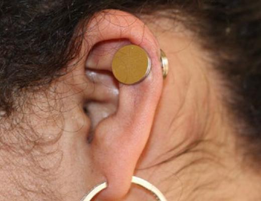
Figure 1. Magnetic disks applied to the upper part of the ear.

Figure 2. Application of silicone gel sheet and magnet in postoperative setting (image courtesy of Dr. Park).
Potential Side effects and Complications
Direct application of magnets to the skin, although well tolerated in most patients, can result in certain complications. Care must be exercised to prevent occurrence of the following potential ill effects.
Contact Dermatitis is a commonly seen complication of metal-based jewelry, often due to an allergic reaction to the alloy. Covering the magnets with a paper tape is a simple method to prevent direct contact between the magnet’s alloy and the skin, thereby preventing an allergic reaction. Park’s method in a post-operative setting is to use silicone gel sheets on both sides of the magnets [5].
Pressure Ulcers may occur due to excess pressure from the magnets [2, 6]. It is always best to use the thinnest magnets (1 mm in thickness) and instruct patients to initially wear only two magnets and remove them if they feel any degree of discomfort. When applied to both sides of the ear, the 1 mm thin magnets will induce a very gentle pressure to the tissue. Patients should be instructed to keep the magnets for as many hours per day as possible, but remove the magnets intermittently: every two hours and allow a 30 minute break before the magnets are re-applied. Magnets should not be worn during sleep.
When two magnets are worn for a few days and are well tolerated, and the ears appear normal and free of dermatitis, open wounds or pressure marks, a third magnet can then be added. The set of three magnets, two on one side and one on the other side, should be worn for a few days to ensure that the pressure applied to the ear tissue is not excessive. If three magnets are well tolerated, a fourth one can be applied to the side with one magnet. No more than four 1 mm thin magnets shall be used as excess pressure will increase the risk of damage to the underlying tissue.

Figure 3. Tissue necrosis at the sites where magnetic disks were applied (image courtesy of Dr. Park).
References
- Ketchum LD, Cohen IK, Masters FW. Hypertrophic scars and keloids. a collective review. Plastic and reconstructive surgery. 1974;53(2):140-154.
- Park TH, Rah DK. Successful eradication of helical rim keloids with surgical excision followed by pressure therapy using a combination of magnets and silicone gel sheeting. International wound journal. 2017;14(2):302-306.
- Gupta S, Jangra RS, Gupta S, Singla R, Use of Neodymium magnetic discs as pressure earrings for ear lobe keloid post-excision, Journal of the American Academy of Dermatology (2018), doi: 10.1016/j.jaad.2018.04.039.
- Tanaydin V, Beugels J, Piatkowski A, et al. Efficacy of custom-made pressure clips for ear keloid treatment after surgical excision. Journal of plastic, reconstructive & aesthetic surgery:jpras. 2016;69(1):115-121.
- Park TH. New pressure device,”Magsil,” as an adjuvant pressure therapy for ear keloids. Arch Facial Plast Surg. 2012 Jul- Aug;14(4):298-9.
- Park TH, Seo SW, Kim JK, Chang CH. Outcomes of surgical excision with pressure therapy using magnets and identification of risk factors for recurrent keloids. Plast Reconstr Surg. 2011 Aug;128(2):431-9.


