KRF Clinical Practice Guidelines in Keloid Disorder (KRF Guidelines®) Treatment Strategy – Version 1.2019
Michael H. Tirgan, MD
INTRODUCTION
This Guideline reviews the overall strategy for treating keloid patients. Readers are advised to consult with site-specific KRF Guidelines that focus on the treatment of particular types of keloids.
It has long been known that successful treatment of a disease is possible only when we understand the underlying pathophysiology. Our current understanding of keloid disorder (KD) is that of a genetic disorder of the wound healing mechanism(s) of the skin, with a highly variable genotype and a very diverse phenotype.
The wound healing defect that leads to the formation of keloid lesions, although not fully understood, perhaps involves the negative feedback loops and signaling pathways that exert control over the wound healing response. Under normal circumstances, once a wound is adequately healed, the inflammatory response, fibroblast proliferation, and collagen production will all regress to their baseline, preinjury status. It is hypothesized that the regression of all normal wound healing responses is under the control of several negative cytokine signaling and feedback mechanisms. Defects in these signaling processes will result in the continuation of the wound healing reaction, excess fibroblast proliferation, and excess collagen deposition, all of which are seen in keloid tissue. This concept is of utmost importance as it can provide the direction for the proper management of keloid patients.
Some de-novo skin pathologies, for instance basal cell carcinoma, can be successfully treated with surgery. However, post-operative recurrence is observed in almost 100% of patients undergoing keloid removal surgery, hence adjuvant ILT or radiation therapy are incorporated to reduce the risk of recurrence.
The core question to ask here is, “Why is there such a high rate of recurrences after keloid removal surgery?” The answer is that keloid removal surgery starts with, and imposes, a totally new wound and a new injury to the skin, thereby triggering the same keloidal wound healing response that produced the original keloid. Perhaps, this is the only medical condition that is triggered, and even made worse, by surgical treatment. It is in this setting that the millennia-old concept of “Primum non nocere – first, do no harm” applies most.
Furthermore, in a situation in which the peak age of onset is in the late teens, one must be mindful of potential harms and side effects of treatment. Therefore, before we expose a large number of young and otherwise healthy individuals to treatments that can be potentially harmful, we need to better understand and to be fully cognizant of all potential short- and long-term side effects of the treatments we are recommending and using.
Focus on keloid patient and not on the keloid lesion
Exposure to radiation therapy and/or frequent injections of high-dose steroids are known to result in long-term, significant, adverse effects. The practice of radiating benign skin conditions was abandoned several decades ago because of the increased incidence of fatal cancers. But once again, we are reverting to radiation therapy as a method to impose surgery to remove keloid lesions, but we are forgetting about the risks to our keloid patients.
Considering the above risks, our strategy should focus on treating the keloid patient as opposed to removing a keloid lesion. We need to focus our attention on reducing the risk of harm to the patients by carefully crafting treatment plans that place the focus on the patient and not on excising a keloid lesion and doing everything we can to prevent recurrence at the surgery site.
Data-driven treatment pathways
The biggest handicap to treating keloid patients is the lack of data-driven treatment pathways. Despite the abundance of patients with all types of keloids, there is a paucity of properly designed and well-conducted clinical trials that can form the foundation for proper clinical management of patients with different types of keloids.
Expert-Driven Treatment Guidelines
Due to the lack of level one evidence for the treatment of keloid lesions, most patients are treated according to the routine and common practices of their treating physicians. This Guidance document also suffers from some of the abovementioned inadequacies; however, the author, to the extent possible, has tried to rely on clinical data and the power of observation and analysis of clinical data from a large cohort of keloid patients. This Guidance document reflects the author’s current thinking, with hopes for future refinement of the recommendations based on validated clinical data.
Proper evaluation of keloid patients
Proper evaluation of a patient starts with a thorough history and examination of the skin. Keloid patients often present for treatment of a particular keloid; however, upon inquiry and examination, it will become evident that most patients have multiple other keloids elsewhere on their skin. History of prior treatment and the response or resistance to all prior interventions should also be elicited.
Patient education
Educating patients about their illness is of utmost importance. Patients need to understand the logic behind how we choose to treat them. Educating patients about the hardships associated with various treatments, expected duration of recovery, frequency and expected side effects, and the cost of treatment will all need to be discussed with the patient before s/he receives our proposed treatments. Patients must make plans to attend treatment sessions and or undergo procedures that require lengthy recovery times. Patient education simply enhances their compliance with treatment.
Setting expectations correctly
Another important matter that must be discussed upfront with all patients is setting realistic expectations from the planned or proposed treatment. Physicians promising a cure or setting high hopes for achieving good results could engender future patient dissatisfaction and non-compliance. Patients who expect perfect results and total clearance of all their keloid lesions in the shortest possible time will quickly be dissatisfied. Thorough and honest discussions must be held between the treating physician and the patient in which realistic expectations are set up front. Such an approach will not only increase patients’ compliance, but will reduce the chances of dissatisfaction with the treatment results.
Setting treatment goals
Treatment strategy starts with establishing the treatment goal, which often varies from patient to patient. A young person with a small facial keloid would most likely desire to see its total disappearance; yet on the other hand, an elderly patient with a post-sternotomy keloid may only desire symptom control. Once the treatment goals are established, patients should be educated about details of each treatment modality that and all potential side effects.
Treatment plans are guided by the morphology of keloid lesions
Current treatment plans are guided by the appearance and to some extent the location of the keloid lesions. This Guidance provides recommendations for the following types of keloid lesions:
- Very early stage disease (papular/linear keloids)
- Nodular / Tumoral Keloids
- Flat Keloids / Keloid Patches
TREATMENT TOOLS
Keloid disorder is one of only a few human illnesses not yet adequately researched. With our limited understanding of its pathophysiology, and the lack of academic and industry interest, most treatment options used to treat keloid patients have been borrowed from other areas of medicine. Current treatment Guidance and KRF recommendations are based on the following methods of treatment.
- Intra-Lesional Triamcinolone (ILT)
- Intra-Lesional Chemotherapy (ILC)
- Contact Cryotherapy
- Pressure devices
At the current time, KRF advises against surgery, radiation therapy, and laser treatment.
KRF’s recommendations are for the most part based on reducing the risk for the iatrogenic worsening of the keloids.
Aggressive treatment of early stage disease
One of the most basic principles of medicine is that treating any illness is easier when the illness is detected at an early stage. The same principle applies to treating keloid patients. It is only with aggressive and non-surgical treatment of early stage keloids that we might be able to gain control over the burden of this disease.
All keloid lesions start as a small papule or a minor liner lesion. The correct approach to such early-stage keloid lesions is to treat them very aggressively with the goal of inducing a complete remission. It is only with this approach that we can have a significant impact on the natural history of this disorder.
For reasons that are beyond this Guidance, quite often a keloid papule does not receive proper treatment and as a result, it grows to becomes a nodule or a small tumor. Often, at that point, a decision is made to remove the lesion surgically. Preventing keloid papules to go down this treatment approach is the cornerstone of successful treatment of keloid patients because allowing a keloid papule to grow and to form a nodule, and removing that nodule surgically, is a path that only leads to the formation of life changing keloids.
Treatment of Keloid Papules
When a keloid papule is first noticed, regardless of its location, it must be dealt with very aggressively and treated with the intention of inducing total regression. Keloid papules must be treated with discipline and followed very carefully. The battle with KD may be lost if the treating physician misses the opportunity to properly treat a keloid papule.
Treating a small keloid papule and forcing it into remission is a lot easier than treating a large keloid lesion. Figure 1 depicts a 31-year-old female with tumoral anterior chest keloid with two adjacent keloid papules at presentation in October 2014. For in-depth history and analysis of this case, please refer to case study #7 in the KRF Guideline for treatment of chest keloids. Figure 2 depicts the same patient in July 2015, after treatment with three cycles of ILT and cryotherapy. What is most noticeable is that two keloid papules were successfully treated yet the main keloid lesion only had a partial response to treatment. Unhappy with the pace of progress, the patient chose to undergo surgery followed by radiation therapy in early 2016. Figure 3 depicts the same patient in October 2017. The papule that was located in the superior margin of the keloid is not visible as it was most likely removed with the surgery; however, despite obvious recurrence of the main keloid, the papule that was present in the inferior aspect of the main keloid, the one that responded to ILT and cryotherapy, remained in remission.
Although it is difficult to predict the fate of these two papules had they not been treated in 2014, what is indisputable is the fact that a durable remission was induced in the lower papule with a combination of ILT and cryotherapy: after close to three years of follow up, there was no keloid regrowth at that site.

Figure 1. A 31-year-old Hispanic female (October 2014) with a tumoral anterior chest keloid. Note the presence of the inflammatory reaction around the margin of the main keloid and multifocal disease with two papular keloids in the immediate vicinity of the main lesion.

Figure 2. The same patient after recovery from three cycles of ILT and cryotherapy (July 2015). Note the thinning of the main keloid along with near- otal flattening of the two keloid papules noticed at presentation.
Algorithm to treat keloid papules
All early-stage keloid papules shall be first treated with ILT (see KRF Guidelines – ILT). Care must be given to accurately inject a low-dose and low-volume of triamcinolone inside the core of the lesion. For lesions that respond to the first injection of ILT, the same treatment shall be repeated once every three to four weeks until maximum response is achieved. If the lesion does not respond to the first injection of ILT, a second injection of ILT shall be attempted.
Lack of response to two ILT injections, or the growth of keloid after the first ILT injection, are grounds to switch the treatment. ILT is a known risk factor for worsening of keloid lesions [1]. The next line of treatment for all such lesions shall be a combination of ILT with contact cryotherapy.
ILC (see KRF Guidelines – ILC) shall be considered for all keloid papules that progress despite ILT and cryotherapy.
Table 1. Summary of treatment recommendations for keloid papules.
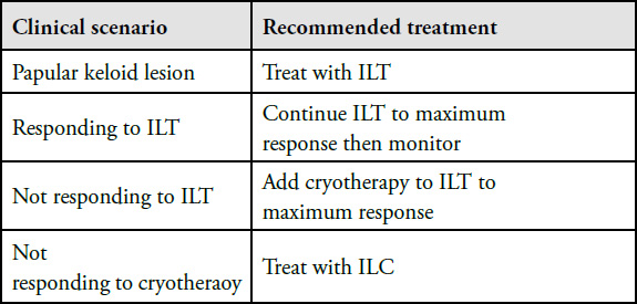
Abbreviations: ILT Intra-lesional triamcinolone. ILC Intralesional Chemotherapy
Figures 3-4 depict the before and after treatment results in 38-year-old Asian patient with multiple anterior chest keloids. Once again, complete remission is achieved in the papular lesions.
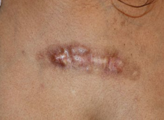
Figure 3. The same patient after surgery and radiation therapy. Note the recurrent disease along the surgical incision line (October 2017). Also, note that the small keloid papule that was previously present under the main keloid lesion, the one that was treated with ILT and cryotherapy, remained in remission for three years.

Figure 4. A 38-year-old Asian male with a thick linear and a few papular anterior chest keloids (Nov 2013).

Figure 5. The same patients, after treatment with ILC (January 2016). Notice overall improvement, most importantly the complete regression of keloid papules in response to ILC.

Figure 6. A 35-year-old Asian female with multifocal keloid disorder involving her face and shoulders with a solitary papule of the anterior chest (June 2013).
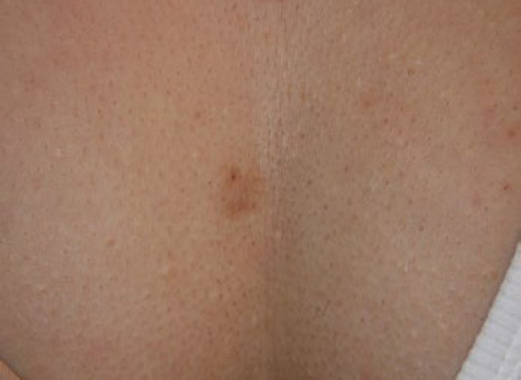
Figure 7. Same patient, April 2014. Note the total regression of the anterior chest keloid in response to treatment with ILT and cryotherapy.

Figure 8. A 36-year-old Caucasian female with multifocal keloid disorder and a history of a few keloid papules on her chest. At presentation, several small papules had previously responded to ILK and lasers, with one particular lesion resisting treatment (September 2013).
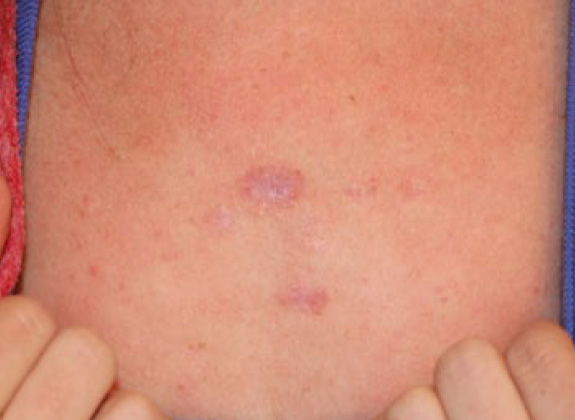
Figure 9. Same patient, December 2015. Note the near-total regression of the anterior chest keloid in response to treatment with ILT, ILC, and cryotherapy.
Making the Case Against Surgery
A 24-year-old African-American male presented in February 2017 with massive neck keloids (Figure 10). His struggles with KD started approximately four to five years earlier when he first noticed several small keloid lesions forming on his upper neck and submental skin. The lesions were first treated with ILT and subsequently removed surgically in early 2015. Recurrent disease was noted soon after surgery and was treated with repeat surgery and post-operative ILT injections. Unfortunately, this approach was not successful and led to the development of massive submental keloids, as shown in Figure 10. The patient was asked to produce any prior images of his neck. Figure 11 depicts the size and location of his neck keloids prior to his first surgery.
As shown in several cases, small papular and nodular keloids can be successfully removed using non-surgical treatments. On the other hand, keloid removal surgery imposes a major risk on all patients undergoing this intervention. Although no one has a crystal ball to predict the outcome of this patient’s neck keloids had he been treated with ILT, ILC, and cryotherapy instead of surgery, patients like this young man should not be denied the chance for non-surgical treatment. Furthermore, surgery is a known cause for the formation of these life changing massive keloids [2,3]. The following two cases presented in Figures 12-15 demonstrate the proof of the principle that small keloid lesions can be successfully treated using non-surgical tools. Several more site-specific examples of the success of this treatment approach can be found within each site-specific Guidance document.
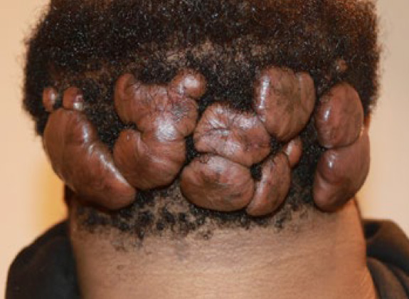
Figure 10. A 24-year-old African American male with multifocal, massive neck keloids (February 2017).

Figure 11. The same patient, December 2014, prior to undergoing surgery. Photographs from patient’s own archives.

Figure 12. Early-stage nodular neck keloid in a 24-year-old African American male (December 2012).
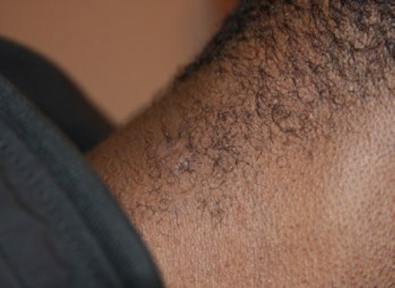
Figure 13. The same patient two years after treatment with cryotherapy (March 2015). Note the minimal scarring at the site of the treated keloid.

Figure 14. A 36-year-old African-American male with submental keloids (May 2016).

Figure 15. The same patient treated with ILK and ILC (June 2018). Note the significant improvement and near total regression of the treated area.
Treatment of nodular / tumoral keloids
Ideally, one wishes for all keloid patients to present at the earliest stage of their disease and to receive effective treatments for their keloid papules. The reality is that many patients present with large and tumoral keloids, either because they have not received any prior treatments, or their keloids have grown despite treatment.
Most tumoral and bulky keloid lesions can be successfully debulked using cryotherapy. Readers are advised to review the site-specific Guidance documents for proper treatment of bulky keloids. Once the mass of a bulky keloid is reduced to the level of the skin, attention must be directed to controlling of the remnant of the keloid tissue that may persist at the base of the keloid.
Treatment of inflammatory and flat keloids
Flat keloids, especially those located on anterior chest skin are among the most difficult keloids to treat. Figure 16 depicts a 34-year-old male who previously underwent two prior surgeries for treatment of anterior chest keloids. Currently, the only approach to keloid lesions like the one shown here is with ILC.
Patients who present with such advanced keloids have often exhausted all available treatment options. Ideally all such patients need a systemic form of treatment that can be taken orally, or intravenously or a highly effective topical product that can be applied to all the diseased areas. Unfortunately, none is currently available.

Figure 16. A 34-year-old Hispanic make with multiple large linear and flat central chest keloids.
Treatment of patients with extensive and multiple keloids
Patients who present with extensive and multiple keloids pose particular challenges as most suffer from a biologically more aggressive form of KD and have a tendency to form numerous keloids at a much faster growth rate compared with patients who only have one or a few keloid lesions. All these patients are in need of a systemic form of treatment, which at the current time does not exist.
With limited effective treatment options, the treating physician together with the patient must prioritize the sites and the lesions that need to be treated and address each particular lesion according to the most appropriate plan of care.
Need for systemic treatments
Many keloid patients suffer from a constantly progressive disease, a situation that cannot be controlled with local treatments. All such patients are in need of systemic treatments. We hope that over time, KD can attract the interest of research community and the critical funding and support needed to explore and to develop totally new treatment frontiers.
Need for research and collaborations
Progress cannot be made unless the medical and the research communities work together with the common goal of better understanding KD’s pathophysiology. This path will hopefully lead to the development of better tools to treat our patients. KRF invites everyone who is interested in researching this condition to join us in tackling this life changing disorder.
REFERENCES
- Tirgan, MH. Intra-lesional Steroid Injections: Can they be harmful to some patients? Results of an online survey. International Journal of Keloid Research. 2017 vol. 1 no. 1, July 1, 21-28.
- Tirgan, MH. Pubic keloids: Evaluation of risk factors and recommendation for keloid staging system, F1000 Research, June 28, 2016.
- Tirgan, MH. Massive ear keloids: Natural history, evaluation of risk factors and recommendation for preventive measures – A retrospective case series, F1000 Research, October 13, 2016.
