
BACKGROUND
Keloids are fibroproliferative disorders characterized by excessive extracellular matrix deposition and progressive growth, yet the neuro-stromal mechanisms driving fibroblast dysregulation remain incompletely understood. Previous studies have well explained the hyperproliferative process during early and active phases of keloids through inflammatory-fibroblast activation and fibrotic signaling pathways such as TGF-β. However, as a chronic, recurrent, and multifactorial disease driven by genetic, immune, mechanical, and hormonal factors, many causes underlying disease progression or recurrence remain elusive.
METHODS
- Single-cell sequencing: 10X Genomics scRNA-seq was performed on human keloid and healthy skin samples, complemented by probe-based 10X Flex for improved detection of low-abundance transcripts.
- Tissue and cell assays: RNAscope, immunofluorescence, qPCR, and immunoblotting were used. Primary fibroblasts were isolated and stimulated with epinephrine/norepinephrine, with or without inhibitors (e.g., metoprolol) or siRNA-mediated knockdown of ADRB1 or TSN.
- Mechanistic studies: cAMP assays, RNA immunoprecipitation (RIP), and chromatin immunoprecipitation (ChIP) were employed to dissect the DRB1–cAMP–PKA–CREBTSN axis.
- Animal model: A rat tail stretched-wound model recapitulating keloid-like pathology was used. Interventions included 6-OHDA (catecholaminergic denervation), Nepicastat (inhibition of catecholamine synthesis), AAV-shAdrb1 (fibroblast-specific Adrb1 knockdown), or oral metoprolol.
RESULTS
1. Osteogenic-like fibroblasts in keloid: A RUNX2⁺ IBSP⁺ fibroblast subpopulation (~25% of dermal cells) was identified in keloid, accompanied by elevated ALP and calcium deposition, indicating hybrid fibro-osseous reprogramming.
2. Aberrant catecholaminergic innervation: Keloid dermis showed dense tyrosine hydroxylase (TH)⁺ nerve fibers, and keloid fibroblasts highly expressed ADRB1.
3. ADRB1-dependent IBSP production: Epinephrine/norepinephrine induced IBSP protein (but not mRNA) in keloid fibroblasts via ADRB1, an effect blocked by metoprolol or ADRB1 knockdown.
4. TSN-mediated translational control: Epinephrine activated the cAMP–PKA–CREB pathway, upregulating TSN transcription. TSN translocated IBSP mRNA from the nucleus to the cytoplasm, enhancing translation. TSN silencing abrogated IBSP production.
5. In vivo validation: Denervation, inhibition of catecholamine synthesis, fibroblast-specific Adrb1 knockdown, or oral metoprolol each reduced scar thickness, collagen deposition, Runx2⁺/Ibsp⁺ cell counts, and GAG accumulation in the rat model.
6. Therapeutic implication: The β1-adrenergic antagonist metoprolol effectively suppressed keloid-like pathology, supporting its repurposing for keloid therapy.
CONCLUSION
Our findings reveal that catecholaminergic nerves directly reprogram fibroblast osteogenic activity via an ADRB1-cAMP-PKACREB-TSN post-transcriptional axis, and provide a mechanistic rationale for repurposing β1-adrenergic antagonists as a targeted therapy for keloid disease.
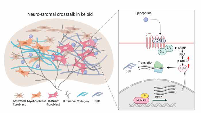
Fig1. Schematic diagram of the mechanism by which neuronal–stromal interactions mediate osteogenic-like pathology in keloids.


